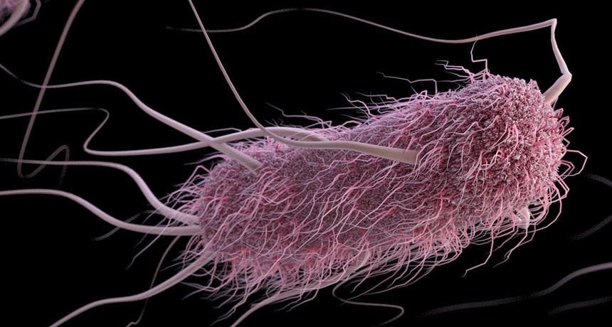
E. coli has a type and it isn’t pretty. The bacterium is more likely to cause severe diarrhea in people with type A blood.
An illness-causing strain of E. coli secretes a protein that gloms onto the sugar molecules that decorate type A blood cells, but not type B or O cells. These sugar molecules also decorate cells lining the intestines of people with type A blood and appear to provide a handle for the bacterium to latch onto before injecting its diarrhea-causing toxins, researchers report May 17 in the Journal of Clinical Investigation. There were hints that blood type was linked to the severity of E. coli infection. But a clear connection was lacking until now, says a team led by researchers at Washington University School of Medicine in St. Louis.
Collaborators at Johns Hopkins University gave 106 healthy volunteers water laced with a strain of E. coli isolated from a person in Bangladesh with severe diarrhea. Within five days, 81 percent of the type A or AB volunteers developed moderate to severe diarrhea compared with roughly half the people with blood types O or B. (Everyone received antibiotics to clear the bacterium).
This discovery suggests that a vaccine targeting the bacterial protein — which is found in many E. coli strains — could be effective. That could help not only travelers but also children in the developing world, where repeated infections are linked to malnutrition and stunted growth. E. coli isn’t the only microbe that can cause severe diarrhea, though, making good hygiene — washing hands and purifying water — still the best defense.
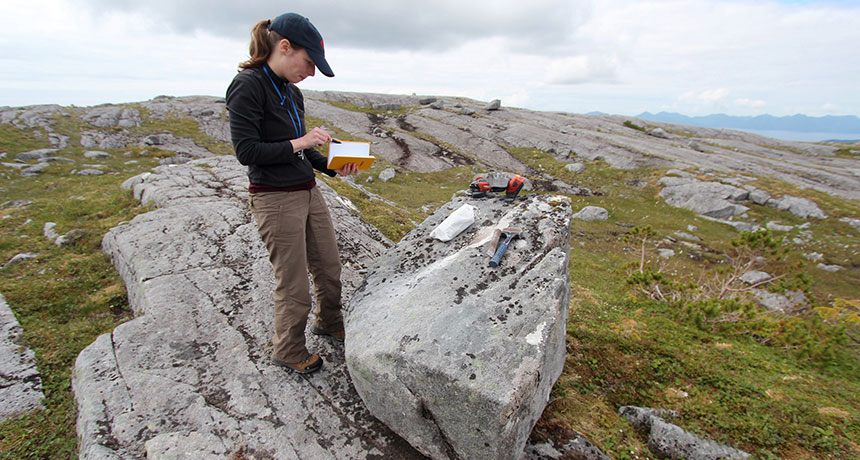
Ancient colonizers of the Americas could have traveled down Alaska’s Pacific coast in canoes or other sea vessels around 17,000 years ago, a new study finds.
At that time, toward the end of the last ice age, glaciers had just receded from a cluster of southern Alaskan islands, say geologist Alia Lesnek of the University at Buffalo in New York and colleagues. Life-supporting habitats appeared soon after the ice melted, the scientists report May 30 in Science Advances. The study is the latest to weigh in on the debate over how humans spread into the New World after arriving from Asia and reaching as far as Florida and South America by 14,500 years ago ( SN: 6/11/16, p. 8 ; SN: 12/26/15, p. 10 ). Previous work has hinted that an inland, ice-free corridor from Alaska through what’s now British Columbia and into the United States may not have contained enough vegetation and wildlife to enable human travel before around 12,600 years ago ( SN Online: 8/10/16 ). New geologic evidence supports the idea of a coastal route, though Lesnek’s team found no human bones or artifacts on the islands. Measures of chemicals that accumulate in rock due to cosmic radiation once glaciers retreat provided age estimates for when four Alaskan islands lost their ice coat. An open pathway for coastal travelers probably existed along the entire southeastern Alaskan coast roughly 17,000 years ago, the scientists say. Radiocarbon dates for a ringed seal’s remains found on a southern Alaskan island indicate that the seal lived about 17,000 years ago, suggesting the area became habitable soon after glaciers left.
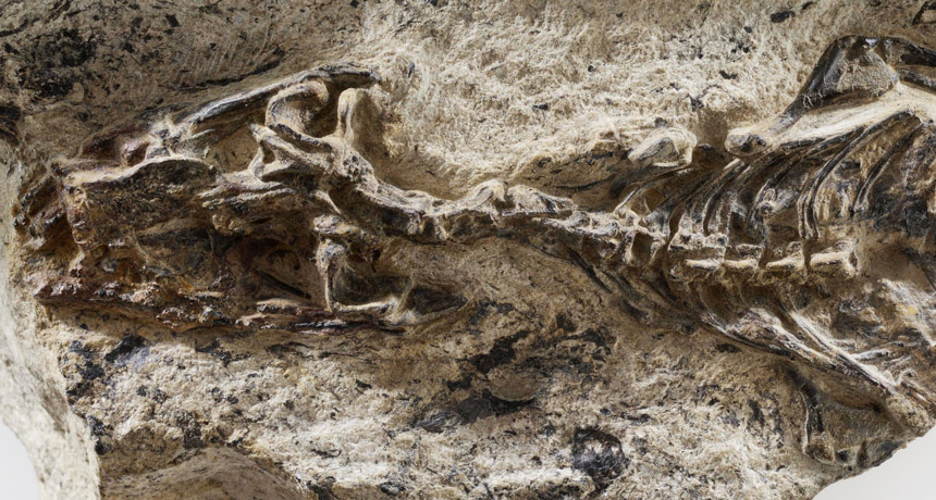
A little animal that washed out to sea 240 million years ago off the coast of what’s now Italy turns out to be the oldest known fossil of a lizard.
The identification pushes back the fossil record of snakes and lizards by about 75 million years, says Tiago Simões of the University of Alberta in Edmonton, Canada. He and colleagues used observations of the fossil, called Megachirella wachtleri, and of related living and extinct species plus genetic data to reconstruct the evolutionary history of squamates, the reptile group that today includes snakes and lizards, the researchers report in the May 31 Nature. “Understanding lizard and snake evolution has been a constant problem in paleontology,” says vertebrate paleontologist Stephanie Pierce of Harvard University who worked on the issue years ago but wasn’t involved in the new study. The trouble comes largely from a lack of relevant fossils, she says. That dearth isn’t just because little animals don’t fossilize as readily as big ones do. “Things like giant dinosaurs — they’re pretty easy to spot,” she says. “But if you’re looking for something that can fit in the palm of your hand, that makes it very challenging.” A collector found M. wachtleri almost 20 years ago in a part of the Italian Alps that had once been underwater. But researchers didn’t categorize it as a full member of the squamate branch of reptiles, until now. The sole specimen is partly embedded in rock, which obscured some of the creature’s telltale features. Now a CT scan has revealed previously unknown squamate details of its palate, braincase, limbs and shoulder, Simões says. M. wachtleri was a small, terrestrial animal with teeth appropriate for snapping up insects, he says. Yet a catastrophe such as a tropical storm apparently swept it out to sea along with masses of leaves.
Besides taking a closer look at M. wachtleri, Simões and some colleagues traveled to 17 countries from Brazil to China to reexamine the other known squamate fossils. Combining all of the new fossil measurements with DNA data, the researchers reconstructed the evolutionary tree of squamates. Among the new insights: Geckos appear to be the most ancient of still-surviving lizards, the team found. And iguanas, a hard-to-place group on the tree, have a more recent origin than some other studies have found.
“The picture is coming together,” says study coauthor Oksana Vernygora, an evolutionary biologist also at the University of Alberta.
Previously, the oldest known squamate fossils dated back about to 168 million years ago. Adding M. wachtleri to the mix means that the squamates are so ancient that they arose before the mass extinction at the end of the Permian Period 252 million years ago, notes herpetologist and evolutionary biologist Jeff Streicher of the Natural History Museum in London who was not involved in the study. That cataclysm came the closest (yet) to wiping out life on Earth. How the ancestors of modern lizards and snakes made it is still a matter of debate.
The spacecraft fire that killed three Apollo astronauts and rocked the space agency a year and a half ago is still being felt.… Last week, after a series of delays … a major milestone was finally reached: the first manned tests of an Apollo spacecraft to include all the new equipment and safeguards incorporated since the fire. — Science News, June 8, 1968.
Update Since that lunar program reboot, hundreds of NASA astronauts have made it to space, and 12 astronauts have walked on the surface of the moon. After NASA retired the U.S. Space Shuttle program in 2011, American astronauts have had to rely on Russia’s Soyuz spacecraft to reach the International Space Station (SN: 6/11/16, p. 4). Once again, U.S. spacecraft, from SpaceX and Boeing, are planned to take American astronauts into space, but delays have pushed those efforts back to late 2019 and 2020.
Stay younger, longer. Great idea. But direct-to-consumer test kits that promise to gauge a person’s biological age by analyzing a drop of blood are not worth the $100 or so investment, says oncologist Mary Armanios. The tests measure the length of telomeres, the bits of DNA that cap and protect the ends of chromosomes. But the consumer tests are unreliable and can be misinterpreted, Armanios says.
“These kinds of tests can do harm, suggesting there is something wrong when there isn’t,” says the Johns Hopkins School of Medicine researcher, who uses a clinical test of telomere length to diagnose and treat people with certain rare disorders. Armanios gets calls from people who panic when they get their results from consumer tests. One man in his 40s was told his telomeres were those of an 80-year-old. He sold his house and quit work to make the most of the short time he was convinced he had left. Worse, she says, because he was under the misguided impression that surgeries shorten telomeres, he had decided to delay removal of a precancerous skin spot.
Armanios trained in the lab of Carol Greider, who shared the 2009 Nobel Prize in physiology or medicine for discovering telomerase, the enzyme that controls telomere lengthening (SN: 10/24/09, p. 14). Today Armanios is clinical director of the telomere center at Johns Hopkins.
There is a wide range of “normal” when it comes to telomere length. Work by her team and others has shown that cells don’t stop dividing or die because of telomere shortening unless the ends get very short, far from the median.
Yet, commercial testing companies will label clients as older than their birthday suggests if their telomeres are anywhere shorter than the median. Longer means younger. But excessively long telomeres are not a guarantee of a long life and may be associated with higher cancer risk.
The test many consumer companies use, quantitative polymerase chain reaction, or qPCR, has a 20 percent variability rate — tests on different days can yield different answers, studies by Armanios and others have shown. The clinical test Armanios’ group uses is flow cytometry and fluorescent in situ hybridization, or flow-FISH, which has a lower, 5 percent, variability rate. The researchers use the more precise test to study a small group of disorders united by telomere defects. For two of the illnesses, treatment decisions can change based on clinical telomere testing. A hereditary form of aplastic anemia, a failure of the bone marrow to make blood cells, can be treated with a stem cell transplant with immune suppression. People whose telomeres are short need less immune suppression with the transplant. In a study by Armanios and colleagues in the March 6 Proceedings of the National Academy of Sciences, telomere testing flagged nine of 38 patients who required a gentler approach. Likewise, patients with short telomeres who are receiving lung transplants for idiopathic pulmonary fibrosis should get low immune suppression. In fact, traditional levels can be lethal. With this knowledge, the transplant can be done more safely.
“The telomere belongs in the clinic,” Armanios says, “and should not be used as a form of molecular palm reading.”
Air pollution caused 3.2 million new cases of diabetes worldwide in 2016, according to a new estimate.
Fine particulate matter, belched out by cars and factories and generated through chemical reactions in the atmosphere, hang around as haze and make air hard to breathe. Air pollution has been linked to chronic conditions such as heart disease and diabetes (SN: 9/30/17, p. 18), but this study is one of the first attempts to quantify the connection for diabetes. Researchers tracked 1.7 million U.S. veterans for almost a decade to assess their risk of developing diabetes. They also used data from global studies on diabetes risk, as well as air quality data from the U.S. Environmental Protection Agency and NASA, to create equations that analyzed the connection between air pollution exposure and diabetes globally.
The new estimate, reported in July in The Lancet Planetary Health, holds air pollution responsible for about 14 percent of new cases of diabetes worldwide. Factors such as genetics, weight, activity level and diet also influence the risk of the disease, which is on the rise globally. (The World Health Organization estimates that 422 million people now live with type 2 diabetes — up from 108 million in 1980.)
The burden isn’t the same around the globe: Unsurprisingly, countries with high pollution levels, such as Pakistan, India and China, also have especially high rates of air pollution-linked diabetes. The United States, which now has comparatively clean air, is also high on the list.
Forget all the ridicule heaped on treadmill-running shrimp.
About 80 percent of U.S. adults think that government spending on medical research, engineering and technology, and basic science usually leads to meaningful advances, a new survey from the Pew Research Center shows. The nonprofit, nonpartisan research organization queried 2,537 people from April 23 to May 6.
No matter where they fell on the political spectrum, a majority of Republicans and Democrats shared that view. Of liberal Democrats surveyed, 92 percent said government investments in basic scientific research “usually pay off in the long run.” Of conservative Republicans, 61 percent agreed. That general agreement broke down when it came to private versus government spending. Two-thirds of conservative Republicans said that private investment alone would be enough to see that scientific progress is made, compared with 22 percent of liberal Democrats.
Surveys in 2017, 2014 and 2009 by Pew also found similar support among Americans for spending taxpayer dollars on science.
Some seemingly silly government-funded experiments, such as the exercising shrimp, have been singled out by politicians and others in the past as examples of wasteful spending. But appearances can be misleading. Putting the crustaceans through their paces, for example, was part of a larger project studying infection in farmed shrimp in hopes of finding treatments.
Today’s young women are more likely to experience depression and anxiety during pregnancy than their mothers were, a generation-spanning survey finds.
From 1990 to 1992, about 17 percent of young pregnant women in southwest England who participated in the study had signs of depressed mood. But the generation that followed, including these women’s daughters and sons’ partners, fared worse. Twenty-five percent of these young women, pregnant in 2012 to 2016, showed signs of depression, researchers report July 13 in JAMA Network Open. “We are talking about a lot of women,” says study coauthor Rebecca Pearson, a psychiatric epidemiologist at Bristol University in England.
Earlier studies also had suggested that depression during and after pregnancy is relatively common (SN: 3/17/18, p. 16). But those studies are dated, Pearson says. “We know very little about the levels of depression and anxiety in new mums today,” she says.
To measure symptoms of depression and anxiety, researchers used the Edinburgh Postnatal Depression Scale — 10 questions, each with a score of 0 to 3, written to reveal risk of depression during and after pregnancy. A combined score of 13 and above signals high levels of symptoms.
From 1990 to 1992, 2,390 women between the ages of 19 and 24 took the survey while pregnant. Of these women, 408 — or 17 percent — scored 13 or higher, indicating worrisome levels of depression or anxiety. When researchers surveyed the second-generation women, including both daughters of the original participants and sons’ partners ages 19 to 24, the numbers were higher. Of 180 women pregnant in 2012 to 2016, 45 of them — or 25 percent — scored 13 or more. It’s not clear whether the findings would be similar for pregnant women who are older than 24 or younger than 19. That generational increase in young women scoring high for symptoms of depression comes in large part from higher scores on questions that indicate anxiety and stress, Pearson says. Today’s pregnant women reported frequent feelings of “unnecessary panic or fear” and “things getting too much,” for instance.
Those findings fit with observations by psychiatrist Anna Glezer of the University of California, San Francisco. “A very significant portion of my patients present with their primary problem as anxiety, as opposed to a low mood,” says Glezer, who has a practice in Burlingame, Calif.
The study’s cutoff score for indicating high depression risk was 13, but Pearson points out that a lower score can signal mild depression. Women who score an 8 or 9 “still aren’t feeling great,” she says. It’s likely that even more pregnant women might have less severe, but still unpleasant symptoms, she says.
The researchers also found that depression moves through families. Daughters of women who were depressed during pregnancy were about three times as likely to be depressed during their own pregnancy than women whose mothers weren’t depressed. That elevated risk “was news to me,” says obstetrician and gynecologist John Keats, who chaired a group of the American College of Obstetricians and Gynecologists that studied maternal mental health. Asking about whether a patient’s mother experienced depression or anxiety while pregnant might help identify women at risk, he says.
Negative effects of stress can be transmitted during pregnancy in ways that scientists are just beginning to understand, and stopping this cycle is important (SN Online: 7/9/18). “You’re not only talking about the effects on a patient and her family, but potential effects on her growing fetus and newborn,” says Keats, of the David Geffen School of Medicine at UCLA.
Although researchers don’t yet know what’s behind the increase, they have some guesses. More mothers work today than in the 1990s, and tougher financial straits push women to work inflexible jobs. More stress, less sleep and more time sitting may contribute to the difference.
Time on social media may also increase feelings of isolation and anxiety, Glezer says. Social media can help new moms get information, but that often comes with “a whole lot of comparisons, judgments and expectations.”
The first global maps of Pluto and its moon Charon are now available, putting a bookend on NASA’s New Horizons mission.
“From a completionist’s point of view, they are all the good data we have, stitched together into a coherent, complete mosaic,” says planetary scientist Ross Beyer of NASA’s Ames Research Center in Moffett Field, Calif.
The charts focus on the 42 percent of Pluto and 45 percent of Charon where New Horizons snapped images from at least two angles during its 2015 flyby, revealing the landscapes’ height and depth (SN: 6/27/15, p. 16). These measurements add topographic detail to already familiar features. For instance, the smooth plains of Pluto’s distinctive, heart-shaped ice sheet, known as Sputnik Planitia, lie two to three kilometers below the region’s rim.
The biggest surprise on Pluto is a 3,200-kilometer-long system of ridges and troughs that traces a single, long line across the dwarf planet. That streak may stretch all the way around the globe, Beyer and colleagues report online June 11 in Icarus. This feature was only visible with all the data put together, Beyer says. The team doesn’t have a good explanation for its formation yet. Charon’s map confirmed that the moon is made up of two large zones: smooth plains in the southern hemisphere and fractured blocks and canyons in the north, Beyer and colleagues report online July 3 in another paper in Icarus (SN: 4/2/16, p. 20). Initially, scientists thought both terrains were at the same elevation, but the new map shows that the plains are situated a kilometer or two lower than the northern terrain. “We don’t know quite why that is yet, but it sure is interesting,” Beyer says.
Other planetary scientists will use these charts to continue unearthing Pluto and Charon’s secrets. “These maps really form the basis, the cartographic foundation, for anything any other scientist will do,” Beyer says.
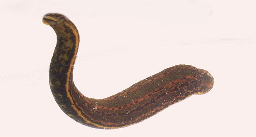
A bacterium found in leeches’ guts needs exposure to only 0.01 micrograms per milliliter of ciprofloxacin to become resistant to that drug, scientists report July 24 in mBio. That’s about 400 times less than the amount of antibiotics thought to trigger drug resistance in this species of bacteria, says study coauthor Joerg Graf, a biologist at the University of Connecticut in Storrs.
Certain leeches are approved for medical use by the U.S. Food and Drug Administration to help patients heal from reconstructive surgery (SN: 10/23/04, p. 266). The slimy creatures suck up blood and secrete anticoagulants, aiding tissue growth. In the early 2000s, researchers noticed an uptick in antibiotic-resistant infections in these patients that were caused by the Aeromonas bacteria found in Hirudo verbana, one of several medicinal leech species. Scientists analyzed the contents of leeches’ stomachs using mass spectrometry, and found drug-resistant bacteria as well as low levels of both ciprofloxacin and enrofloxacin, a veterinary antibiotic used on poultry farms. The researchers say the leeches may have been exposed to these antibiotics through poultry blood used for food on leech farms.
Graf suggests that leech farmers eliminate ciprofloxacin and other antibiotics from their operations. But Aeromonas is also found in freshwater environments. “It is concerning because similarly low amounts [of antibiotics] have been detected in the environment,” he says.
It’s unclear if Aeromonas alone has this lower drug resistance threshold, or if other bacteria can also become resistant at a lower threshold. If so, that could complicate global efforts to prevent drug-resistant infections.